Power Pitch
Pitch: MRE, Diffusion & APT for Body Applications
ISMRM & ISMRT Annual Meeting & Exhibition • 04-09 May 2024 • Singapore

Power Pitch
Pitch: MRE, Diffusion & APT for Body Applications
| 13:45 |
0192.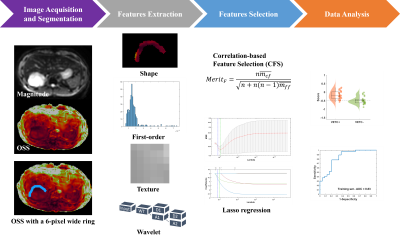 |
Predicting VETC in Hepatocellular Carcinoma Using Radiomic
Features from Peritumoral Mechanical Strain Assessed by MR
Elastography
Keni Zheng1,
Mengsi Li2,
Yi Sui1,
Xiang Shan1,
Emi Hojo1,
Armando Manduca3,
Richard Ehman1,
Jin Wang2,
and Ziying Yin1
1Radiology, Mayo Clinic, Rochester, MN, United States, 2The Third Affiliated Hospital of Sun Yat-Sen University, Guangzhou, China, 3Physiology and Biomedical Engineering, Mayo Clinic, Rochester, MN, United States Keywords: Liver, Liver, MRI, MR Elastography (MRE), HCC, VETC, Radiomics, texture features Motivation: Vessels encapsulating tumor clusters (VETC) is a powerful indicator of aggressive Hepatocellular carcinoma (HCC) associated with recurrence, which is expected to be noninvasively identified using imaging techniques. Goal(s): This study aims to develop a potential mechanical biomarker utilizing radiomics features extracted from MR Elastography (MRE)-assesed peritumoral shear strain to predict VETC in HCC. Approach: Radiomics features were extracted from the tumor boundary on an octahedral shear strain (OSS) map. Feature selection techniques were used to identify relevant features to construct a radiomics score. Results: Three radiomics features were utilized to construct a radiomics score, demonstrating potential for MRE-based strain analysis in predicting VETC. Impact: A radiomics score constructed by radiomics features derived from MRE-based strain analysis in the peritumoral region show promise in predicting VETC in HCC patients. This study results show non-invasive VETC prediction potential in HCC, impacting personalized treatment and patient outcomes. |
| 13:45 |
0193.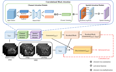 |
Multi-b values DWI-based Virtual High-resolution MR Elastography
and Validation on Liver Fibrosis and Inflammation Prediction
Longyu Sun1,
Yikun Wang2,
Xumei Hu1,
Xueqin Xia3,
Mengting Sun1,
Qing Li1,
Meng Liu1,
Yinghua Chu4,
Xinyu Zhang1,
Ruokun Li2,
and Chengyan Wang1
1Human Phenome Institute, Fudan university, Shanghai, China, 2Shanghai Jiao Tong University School of Medicine, Shanghai, China, 3Institute of Science and Technology for Brain-Inspired Intelligence, Fudan University, Shanghai, China, 4Simens Healthineers Ltd, Shanghai, China Keywords: Liver, Liver, DWI, MRE, Liver fibrosis, RegGAN, CBAM Motivation: Liver biopsy is the standard clinical approach for diagnosing liver fibrosis. However, its invasiveness and possibility of sampling errors impose inherent limitations. Goal(s): To evaluate the reproducibility and reliability of the virtual MRE based on DWI and compare it with MRE to assess its efficiency in diagnosing liver fibrosis. Approach: RegGAN was employed to forecast MRE and rectify the outcomes. And CBAM was utilized to quantify the influence of diverse b-values in DWI. Results: RegGAN-CBAM demonstrates favorable performance in both image and stiffness prediction of MRE. The liver fibrosis grading based on stiffness predictions demonstrates a high level of accuracy and sensitivity. Impact: The prediction methodology of 3D MRE based on DWI exhibits a notable level of diagnostic efficiency and reliability. This approach serves as a non-invasive method with practical applicability in the clinical assessment of liver fibrosis. |
| 13:45 |
0194.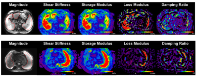 |
Three-dimensional MR Elastography Identifies Portal Hypertension
in Cirrhosis: A Prospective Multicenter Study
Zhiying Wang1,
Minghui Zhou1,
Chen Pan1,
Baihe Luo1,
Jialin Li1,
Qiang Liu1,
and Yu Shi1
1Department of Radiology, Shengjing Hospital of China Medical University, Shenyang, Liaoning, China Keywords: Liver, Elastography Motivation: Hepatic venous pressure gradient (HVPG) is the gold standard for diagnosing portal hypertension, but it is invasiveness, cost, and feasibility. Goal(s): To develop a non-invasive model based on Three-dimensional (3D) MR elastography (3D-MRE) to detect portal hypertension.
Approach: Spearman correlation analysis between
3D-MRE parameters and HVPG; Multivariable linear regression
analysis between mechanical parameters and HVPG; Logistic
regression analysis and establish a model to diagnose
portal hypertension. Results: 3D-MRE is a non-invasive, rapid, and highly accurate tool for predicting portal hypertension. Especially Spleen stiffness at 60Hz was the independent parameters associating HVPG. Impact: This indicates that the use of 3D-MRE can provide more personalized evaluation and better medical experience for patients with cirrhosis and provides a new method for non-invasive diagnosis of portal hypertension. |
| 13:45 |
0195.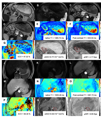 |
Baseline virtual MR elastography and extracellular volume
fraction in the prediction of response to neoadjuvant
chemotherapy in gastric cancer
Yongjian Zhu1,
Wei Cai1,
Yueluan Jiang2,
and Liming Jiang1
1Department of Diagnostic Radiology, National Cancer Center/National Clinical Research Center for Cancer/Cancer Hospital, Chinese Academy of Medical Sciences and Peking Union Medical College, Beijing, China, 2MR Research Collaboration, Siemens Healthineers, Beijing, China Keywords: Digestive, Elastography, gastrointestinal; Cancer Motivation: Although neoadjuvant chemotherapy (NAC) was recommended for gastric cancer (GC), only about 40% patients could achieve pathological response. Accurate prediction of the NAC response is crucial for patients’ benefits. Goal(s): To evaluate the predictive performance of virtual MR elastography (vMRE) and extracellular volume (ECV) for predicting the response to of NAC in GC patients. Approach: Patients underwent DWI-based elastography, pre- and post-contrast T1 mapping before treatment. DWI-based virtual shear modulus (μDiff) and ECV were calculated from DWI and T1 mapping. Results: Both μDiff (AUC: 0.833) and ECV (AUC: 0.794) were independent predictors for NAC response, their combination could improve the AUC to 0.968. Impact: Our result revealed that DWI-based virtual shear modulus (μDiff) and ECV exhibited a promising predictive ability for predicting response to NAC. This would aid in identifying responders before treatment, reducing unnecessary toxicity and side effects, and guiding individualized treatment. |
| 13:45 |
0196. |
Free-breathing Motion-Corrected Liver DWI at 3T with Optimized
Fat-suppression Scheme
Zhiyong Chen1,
Zhangli Xing2,
Enshuang Zheng1,
Mingcong Luo2,
Caixia Fu3,
Guijin Li4,
Thomas Benkert5,
Yunjing Xue1,
and Bin Sun1
1Radiology, Fujian Medical University Union Hospital, Fuzhou, China, 2Fujian Medical University Union Hospital, Fuzhou, China, 3MR Application Development, Siemens Shenzhen magnetic Resonance Ltd., Shenzhen, China, Shenzhen, China, 4MR application, Siemens Healthineers Ltd,Guangzhou,China, Guangzhou, China, 5MR Application Predevelopment, Siemens Healthineers AG, Erlangen, Germany, Erlangen, Germany Keywords: Liver, Diffusion/other diffusion imaging techniques Motivation: A free-breathing motion-insensitive and high-quality DWI is desired for abdominal DWI in clinical practice. Goal(s): This study is to propose and evaluate a novel ss-EPI DWI sequence incorporating motion correction , complex averaging , and a combination of a reparametrized sinc fatsat pulse with an optimized water excitation pulse for abdominal DWI. Approach: Four different DWI sequecnes were performed on volunteers. The overall image qualities and the SNR on the liver papernchyma were evaluated and compared. Results: The SNRs of the liver and the image qualities were significantly higher with the proposed Free-breathing motion-corrected DWI ( MOCO-DWI ) sequence compared to other EPI DWI sequences. Impact: The free-breathing MOCO-DWI sequences with complex-averaging and new fat suppression scheme is recommended for high-quality liver DWI in clinical routine. |
| 13:45 |
0197. |
Intravoxel incoherent motion diffusion-weighted imaging
(IVIM-DWI) of pancreas for assessment of β-cell dysfunction in
hyperglycemia
Ping Liu1,
Yingying Song2,
Wanyi Zhen1,
and Guihua Jiang1
1Department of Medical Imaging,, Guangdong Second Provincial General Hospital, Guangzhou, China, 2Department of Radiology, Affiliated Hospital of Jianghan University, Wuhan, China Keywords: Endocrine, Pancreas, Type 2 diabetes mellitus Motivation: Early detection of damaged β-cell function may help timely protect and stop the progression of hyperglycemia to type 2 diabetes mellitus (T2DM). The impaired β-cell function may be associated with damaged pancreatic microstructure. Goal(s): The pancreatic microstructural changes may serve as the biomarker for β-cell dysfunction. Approach: We evaluate the microstructural changes of the pancreas in patients with hyperglycemic employing intravoxel incoherent motion diffusion-weighted imaging (IVIM-DWI) and explore its correlation with the β-cell function. Results: IVIM-DWI can effectively distinguish T2DM from hyperglycemia, it has the potential for identifying damaged b-cell function for patients with early-stage hyperglycemia but without obvious clinical manifestation of DM. Impact: IVIM-DWI of pancreases is a reliable and non-invasive tool with great potential in detecting the early damaged β-cell function when the DM is still in the insidious stage, and help for improving diabetes diagnosis and management. |
| 13:45 |
0198. |
Abdominal Foundation Model: Bootstrapping artificial
intelligence for MRI organ volume biomarker analysis in ADPKD
Chenglin Zhu1,
Xinzi He2,
Zhongxiu Hu1,
Hreedi Dev1,
Dominick J. Romano1,
Arman Sharbatdaran1,
Anna Prince1,
Andrea Soto Figueroa1,
Sophie J. Wang1,
Hui Yi Ng He1,
Jon D. Blumenfeld3,
and Martin R. Prince1,4
1Weill Cornell Medicine, New York City, NY, United States, 2Cornell University and Cornell Tech, New York City, NY, United States, 3The Rogosin Institute, New York City, NY, United States, 4Columbia University Vagelos Collage of Physicians and Surgeons, New York City, NY, United States Keywords: Kidney, Segmentation, ADPKD Motivation: Abdominal organ volumes are critical MRI biomarkers in many diseases including autosomal dominant polycystic kidney disease. Goal(s): We aim to develop a segmentation model with an enhanced ability to generalize across various abdominal organs and MR pulse sequences. Approach: We construct a multi-modality abdominal foundation model expanding upon our existing ADPKD kidney model which adapts to diverse organs and tissues with minimal new training data. Results: The model was trained using a model-in-loop methodology and evaluated against radiologist benchmarks, yielding an impressive Dice score of 0.94 for in-distribution sequences and 0.73 for organ segmentations on out-of-distribution sequences. Impact: This foundational model can seamlessly integrate into clinical workflows, utilizing routine cases to enhance its performance and extending its application to additional organs and tissues. This advance also marks a significant step toward the automation of MRI reporting. |
13:45 |
0199. |
Water/fat separated Prostate Diffusion-Weighted Imaging using
Dixon-encoded multi-shot EPI with structured low-rank
reconstruction
Yiming Dong1,
David Atkinson2,
Kirsten Koolstra3,
Matthias J.P. van Osch1,
and Peter Börnert1,4
1C.J. Gorter MRI Center, Department of Radiology, LUMC, Leiden, Netherlands, 2Centre for Medical Imaging, University College London, London, United Kingdom, 3Philips, Best, Netherlands, 4Philips Research Hamburg, Hamburg, Germany Keywords: Prostate, Prostate Motivation: Conventional single-shot EPI (ssh-EPI) is fast, but often causes geometric distortions, especially near the rectum. Goal(s): In this research, a Dixon-msh-EPI technique is validated for prostate Diffusion-weighted Imaging (DWI). Approach: Dixon-msh-EPI is proposed to reduce such distortions, while jointly separating water/fat components by a special structured low-rank reconstruction that also corrects shot-to-shot phase variations. Results: Experiments were conducted on 7 healthy male volunteers using a 3T scanner comparing DW-ssh-EPI and Dixon-msh-EPI performance. The Dixon-msh showed significantly reduced geometric distortion and effective fat signal suppression, as scored by two readers. While both methods provided comparable image quality, Dixon-msh-EPI demonstrated improved motion-correction and geometric accuracy. Impact: Dixon-msh-EPI offers improved prostate DWI by significantly reducing geometric distortions and enhancing across b-value registration. This technique could lead to more accurate diagnoses and has the potential to refine clinical MRI protocols, emphasizing precision in prostate diffusion imaging. |
| 13:45 |
0200.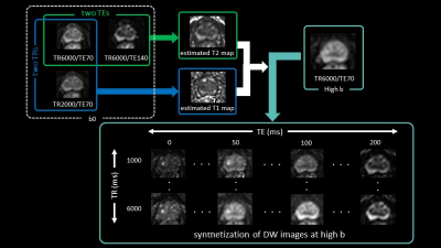 |
Value of quantitative relaxation mapping calculated from
Multiple-Repetition Multiple-Echo DWI acquisition in prostate
cancer detection
Tsutomu Tamada1,
Yu Ueda2,
Mitsuru Takeuchi3,
Atsushi Higaki4,
Yuichi Kojima4,
Yoshihiko Fukukura4,
and Akira Yamamoto4
1Radiology, Kawasaki Medical School, Kurashiki, Japan, 2Philips Japan, Tokyo, Japan, 3Radiolonet Tokai, Nagoya, Japan, 4Kawasaki Medical School, Kurashiki, Japan Keywords: Prostate, Prostate Motivation: The detection rate of csPCa in PI-RADS 3 lesions is only 25-38%. This leads to unnecessary biopsies. Goal(s): Can multiparametric quantitative maps based on estimated T1 and T2 help differentiate clinically significant prostate cancer (csPCa) from non-csPCa in PI-RADS 3 lesions? Approach: We compare T1, T2, and ADC obtained from Multiple-Repetition time Multiple- Echo time (MRME) based DWI (MRME-DWI) between csPCas and non-csPCas in PI-RADS 3 lesion using MRI-ultrasound fusion targeted biopsy as the reference standard. Results: The T1, T2, and ADC were significantly lower for csPCas than non-csPCas. Combining these indices yields an AUC of 0.82. Impact: MRME-DWI can simultaneously obtain T1, T2, and ADC from a single region of interest in a single imaging session. Combining these quantitative measures is expected to improve the detection rate of clinically significant prostate cancer in PI-RADS 3 lesions. |
| 13:45 |
0201.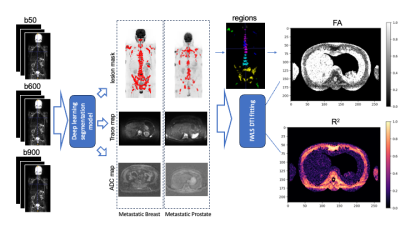 |
Estimating diffusion fractional anisotropy of disease in
patients with advanced prostate and breast cancers. A
single-center study.
Sheng Yu1,
Antonio Candito1,
Konstantinos Zormpas-Petridis 1,2,
Ana Ribeiro1,3,
Georgina Hopkinson3,
Jessica M Winfield1,3,
Nina Tunariu1,3,
Christina Messiou1,3,
Dow-Mu Koh1,3,
and Matthew David Blackledge1
1Institute of Cancer Research, London, United Kingdom, 2Fondazione Policlinico Universitario Agostino Gemelli, Rome, Italy, 3The Royal Marsden NHS Foundation Trust, London, United Kingdom Keywords: Cancer, Whole Body, Metastasis, Fractional Anisotropy
Motivation: Whole-body diffusion-weighted imaging in
oncology assumes isotropic diffusion therefore that data
probing the directional dependence of diffusion in body
cancer applications is scarce.
Goal(s): To report the distribution of fractional
anisotropy (FA) values in metastatic bone disease for a
cohort of patients who underwent multi-directional diffusion
weighted imaging.
Approach: Deep learning was used to auto-segment
metastatic lesions; FA values are calculated for each
segmented region and the distributions reported.
Results: FA distributions from 85 patients with
metastatic disease (50 prostate, 33 breast) are reported and
are deemed to be low (mean FA range breast group: 0.2179 -
0.3132; prostate group: 0.2866 - 0.3717). Impact: We have conducted diffusion tensor imaging analysis for whole body DWI data and carried out the first bone lesion FA comparison study. This work provides a rationale for choice of diffusion-weighting gradient schemes in clinical imaging. |
| 13:45 |
0202.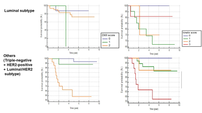 |
Predicting Disease-Free Survival by DWI and DCE MRI Scores for
Breast Cancer Patients with Neoadjuvant Systemic Treatment
Rie Ota1,
Masako Kataoka2,
Mami Iima2,
Maya Honda3,
Aika Okazawa2,
Mizue Suzuki1,
Shotaro Kanao1,
Takeshi Kubo1,
Yosuke Yamada4,
Yasuhide Takeuchi4,
Masahiro Takada5,
and Yuji Nakamoto2
1Department of Radiology, Tenri Hospital, Nara, Japan, 2Department of Diagnostic Imaging and Nuclear Medicine, Kyoto University graduate school of medicine, Kyoto, Japan, 3Department of Diagnostic Radiology, Kansai Electric Power Hospital, Osaka, Japan, 4Department of Diagnostic Pathology, Kyoto University Hospital, Kyoto, Japan, 5Department of Breast Surgery, Kyoto University Hospital, Kyoto, Japan Keywords: Breast, Breast Motivation: MRI is expected to be a new surrogate marker of prognosis that can replace pathological complete response (pCR) for breast cancer patients who underwent neoadjuvant systemic treatment (NST). Goal(s): MRI is expected to be a new surrogate marker of prognosis that can replace pathological complete response (pCR) for breast cancer patients who underwent neoadjuvant systemic treatment (NST) Approach: Survival analysis was performed for disease-free survival using Kaplan-Meier method. Results: DWI score after NST of breast cancer was associated with DFS, in particular triple-negative, HER2-positive, and luminal-HER2 subtype. Impact: The DWI / kinetic score obtained from MRI after neoadjuvant systemic treatment (NST) were associated with disease-free survival (DFS), especially among triple-negative, HER2-positive, and luminal/HER2 subtype. The DWI score may be a biomarker for prognosis of breast cancer patients. |
| 13:45 |
0203.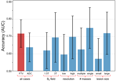 |
Apparent diffusion coefficient as a non-contrast marker of
residual disease after breast neoadjuvant treatment
Patrick J Bolan1,
Wen Li2,
Bonnie N Joe2,
Nu Le2,
Elissa Price2,
Jessica Gibbs2,
Lisa J Wilmes2,
Debsmita Biswas3,
Anum Kazerouni3,
An L Church4,
Elizabeth S McDonald5,
Stephane Loubrie6,
Rebecca Rakow-Penner6,
Hon J Yu6,
Dariya Malyarenko7,
Thomas L Chenervert7,
Beatriu Reig8,
Nola M Hylton2,
and Savannah Partridge3
1Center for MR Research / Radiology, University of Minnesota, MINNEAPOLIS, MN, United States, 2Radiology, University of California San Francisco, San Francisco, CA, United States, 3University of Washington, Seattle, WA, United States, 4Radiology, University of Minnesota, MINNEAPOLIS, MN, United States, 5Radiology, University of Pennsylvania, Philadelphia, PA, United States, 6Radiology, University of Californa San Diego, San Diego, CA, United States, 7Radiology, University of Michigan, Ann Arbor, MI, United States, 8Radiology, New York University, New York, NY, United States Keywords: Breast, Treatment, Cancer, Treatment Response Motivation: Accurate imaging markers to establish pathologic complete response (pCR) during neoadjuvant chemotherapy (NACT) could enable therapy de-escalation to avoid excessive systemic treatments Goal(s): To determine if quantitative diffusion-weighted MRI (DWI) can accurately detect pCR following NACT. Approach: In the ACRIN 6698/I-SPY 2 multicenter trial dataset, tumor region apparent diffusion coefficient (ADC) from DWI was measured on post-NACT/presurgical MRIs. The accuracy of ADC for predicting pCR, alone and in combination with functional tumor volume (FTV) from contrast-enhanced MRI, was assessed. Results: In multivariate models ADC accurately predicts pCR, and is influenced by field strength, spatial resolution, and lesion morphology. Impact: Apparent diffusion coefficient measured by DWI shows promise for determining absence of residual disease following chemotherapy and may provide a non-contrast option for tailoring therapies and enabling patients to avoid unnecessary prolongation of treatment. |
| 13:45 |
0204.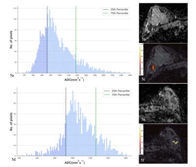 |
Multiparametric MRI-based radiomics fusion combined with
quantitative stratified ADC-defined tumor habitats for TNBC
identification
Wanli Zhang1,2,
Fangrong Liang1,2,
Jiamin Li1,2,
Yongzhou Xu3,
Xin Zhen4,
Ruimeng Yang1,2,
and Xinqing Jiang1,2
1Department of Radiology, The Second Affiliated Hospital, School of Medicine, South China University of Technology, Guangzhou, China, 2Department of Radiology, Guangzhou First People's Hospital, Guangzhou, China, 3Philips Healthcare, Guangzhou, China, 4School of Biomedical Engineering, Southern Medical University, Guangzhou, China Keywords: Breast, Cancer, Breast cancer, MRI, Triple-negative breast cancer, Tumor habitat Motivation: Investigated the role of quantitative stratified apparent diffusion coefficient (ADC)-defined tumor habitats in differentiating triple-negative breast cancer (TNBC) from non-TNBC using a multiparametric MRI (mpMRI)-based feature fusion radiomics (RFF) approach. Goal(s): To develop an RFF-StratifiedADC model using an RFF strategy and reveal distinct ADC map–based tumor habitats for distinguishing TNBC. Approach: RFF (predominant MRI sequence–based fused features), RADC (ADC radiomics features), StratifiedADC (stratified ADC–defined tumor habitat parameters), and combined RFF-StratifiedADC models were constructed to identify TNBC. Results: Stratified ADC parameters helped evaluate the underlying biological proliferation and cellularity within tumor habitats. The integrated RFF-StratifiedADC model was effective and reliable for TNBC identification. Impact: Stratified ADC–defined tumor habitat parameters derived from whole-tumor ADC maps, along with fused radiomics features from dominant mpMRI sequences (T2WI, DWI, ADC maps, and DCE2), can serve as potential biomarkers for differentiating TNBC from non-TNBC. |
| 13:45 |
0205.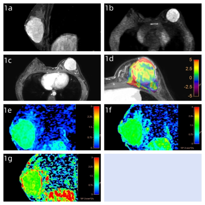 |
Amide-proton transfer weighted imaging combined with
time-dependent diffusion weighted imaging for evaluation of
breast tumors
Xiaoyan Wang1,
Yan Zhang1,
Jingliang Cheng1,
Liangjie Lin2,
Zhigang Wu2,
Peng Sun2,
Ying Hu1,
Anfei Wang1,
Ruhua Wang1,
Yong Zhang1,
Ying Li1,
Kun Zhang1,
and Wenhua Zhang1
1The First Affiliated Hospital of Zhengzhou University, Zhengzhou, China, 2Philips Healthcare, Beijing, China Keywords: Breast, Breast Motivation: Breast cancer is a complex and heterogeneous disease, and its different subtypes show very different biological characteristics, which will also affect the choice of treatment. Goal(s): The objective of this study was to explore the value of APTWI combined with td-MRI in evalution of breast tumors. Approach: The performance of APTWI was compared to conventional td-MRI in differentiation between benign and malignant breast tumors. Results: APTWI overperform td-MRI in differentiation between benign and malignant breast tumors. And ADC values derived from td-MRI at different gradient oscillation frequencies show potential diagnostic values in predicting different risk factors in breast cancer. Impact: These may provide new ideals for the diagnosis, treatment and prognosis evaluation of breast cancer. |
| 13:45 |
0206.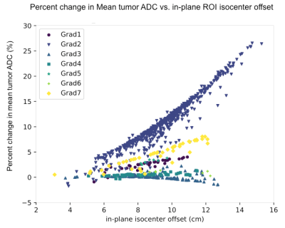 |
Effects of multi-vendor gradient non-linearity correction on
breast tumor ADC measurements in the ACRIN 6698 trial
Lisa J Wilmes1,
Wen Li1,
Judith Zimmermann1,2,
David C Newitt1,
Dariya I Malyarenko3,
Jiachao Liang1,
Patrick J Bolan4,
Savannah C Partridge5,
I-SPY 2 Investigator Network6,
Thomas L Chenevert7,
and Nola M Hylton1
1Radiology and Biomedical Imaging, University of California San Francisco, San Francisco, CA, United States, 2Radiology, Stanford University, Stanford, CA, United States, 3Radiology, University of Michigan, Ann Arbor, MI, United States, 4Radiology, University of Minnesota, Minneapolis, MN, United States, 5Radiology, University of Washington, Seattle, WA, United States, 6Quantum Leap Healthcare, San Francisco, CA, United States, 7University of Michigan, Ann Arbor, MI, United States Keywords: Breast, Diffusion/other diffusion imaging techniques, gradient nonlinearity correction, breast cancer, ADC Motivation: MRI scanner gradient non-linearity (GNL) is a known source of variability and spatially-dependent bias in quantitative ADC measurements derived from diffusion-weighted imaging (DWI). Goal(s): Evaluate the effects of GNL correction on DWI from the ACRIN 6698 multi-center trial, which investigated ADC as a marker breast cancer response in patients receiving neoadjuvant therapy. Approach: Retrospective GNL correction was performed on the ACRIN 6698 DWI data Results: Percent decrease in mean tumor ADC post-GNC ranged from 0.5%-11% for different MRI gradient sets, illustrating that GNL can confer significant bias to ADC measurements and should be corrected in multi-center clinical DWI trials. Impact: Implementation of gradient non-linearity correction to reduce bias and variability of tumor ADC measurements from DWI data acquired in multi-center, multi-platform oncology trials may improve the quantitative utility of ADC for characterizing response to treatment. |
| 13:45 |
0207.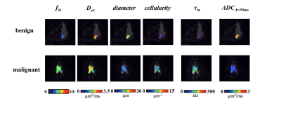 |
The diagnostic performance of time-dependent diffusion MRI in
differentiating benign and malignant breast tumors
Jie Lu1,
Xue Li2,3,
Haotian Li1,
Kuiyuan Liu1,
Chunmei Li2,3,
Min Chen2,3,
and Dan Wu1
1Key Laboratory for Biomedical Engineering of Ministry of Education, Department of Biomedical Engineering, College of Biomedical Engineering & Instrument Science, Zhejiang University, Hangzhou, China, 2Department of Radiology, Beijing Hospital, National Center of Gerontology, Institute of Geriatric Medicine, Chinese Academy of Medical Sciences, Beijing, China, 3Graduate School of Peking Union Medical College, Chinese Academy of Medical Sciences, Beijing, China Keywords: Breast, Tumor, diffusion-time-dependence Motivation: Diffusion-time-dependent diffusion MRI (td-dMRI) has potential in noninvasive mapping of breast tumor microstructure. However, its diagnostic value in differentiating benign and malignant tumors remains unclear, especially including transcytolemmal water exchange. Goal(s): To investigate the clinical value of td-dMRI-based microstructural mapping for discriminating benign and malignant breast tumors. Approach: Time-dependent dMRI data acquired with OGSE and PGSE sequences were estimated with the JOINT models. Results: The td-dMRI-based fin, cellularity and τin showed significant group differences between benign and malignant breast tumors. The combination of cellularity and τin indices achieved the highest diagnostic performance, with an accuracy of 94% and AUC of 0.958. Impact: The diagnostic performance of cellularity-τin obtained from td-dMRI was comparable with or superior to previous studies. The 7.5-minute protocol is translatable to clinical diagnostics. |
| 13:45 |
0208.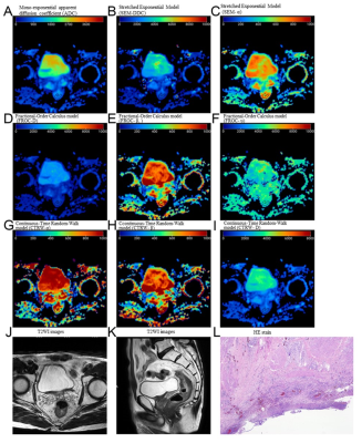 |
Predictive value of mono-exponential and mathematical models in
rectal cancer responsiveness to neoadjuvant chemoradiotherapy
Mi Zhou1,
Meining Chen2,
Qin Zhang3,
and Longlin Yin1
1Department of Radiology, Sichuan Provincial People's Hospital, University of Electronic Science and Technology of China, Chengdu, China, 2MR Research Collaboration, Siemens Healthineers, Chengdu, China, 3MRI clinical application, Customer Service Department, Siemens Digital Medical Technology Co., LTD, Shanghai, China Keywords: Treatment Response, Treatment Motivation: Although it is challenging to predict LARC responsiveness to nCRT, the potential of emerging non-Gaussian DWI models for this purpose remains unexplored. Goal(s): To assess the efficacies of mono-exponential ADC and various non-Gaussian DWI models, including SEM, FROC, and CTRW, in predicting LARC responsiveness to nCRT. Approach: This prospective study included 103 LARC patients. Various DWI models were assessed, and post-surgery histopathology was utilized to classify patients based on responsiveness to nCRT.
Results: Non-Gaussian models, especially CTRW
parameters, demonstrated robust capacity to predict both pCR
and T-downstaging. The combination of CTRW parameters
yielded the best diagnostic performance. Impact: This study demonstrated the potential for novel non-Gaussian DWI models to enhance predictions of LARC responsiveness to nCRT, facilitating optimized treatment plans and encouraging further research in precision oncology. |
| 13:45 |
0209.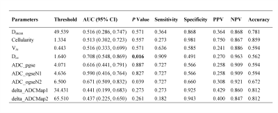 |
Time-dependent Magnetic Resonance Imaging for Predicting
Pathological Complete Response in Rectal Cancer with Neoadjuvant
Therapy
Xiaoling Gong1,
Yu Shen2,
Xiaoxiao Zhang3,
Bing Wu1,
and Bin Song1
1Departments of Radiology, West China Hospital, Sichuan University, Guoxue Xiang No. 37, Chengdu, Sichuan, 610041, PR China, Chengdu, China, 2Colorectal Cancer Center, Department of General Surgery, West China Hospital, Sichuan University, Chengdu, 610041, People’s Republic of China, Chengdu, China, 3Department of Clinical, Philips Healthcare, China, Wuhan, China Keywords: Pelvis, Diffusion/other diffusion imaging techniques, rectal cancer;neoadjuvant treatment;pathological complete response;oscillatory gradient spin echo Motivation: Time-dependent (TD) magnetic resonance imaging (MRI), an emerging imaging modality, offers insights into cellular microstructures, but its relevance to rectal cancer's neoadjuvant treatment response remains unknown. Goal(s): To determine the value of TD MRI-derived metrics for preoperatively predicting pathological complete response (pCR) in rectal cancer. Approach: Univariate and multivariate logistic regression analyses were used to identify key predictors of pCR. Results: Extracellular diffusivity independently predicted pCR, with a cutoff of 1.640 effectively distinguishing between pCR and non-pCR tumors. Impact: Time-dependent magnetic resonance imaging emerges as a promising tool for detecting the pathological complete response in rectal cancer following neoadjuvant therapy, potentially facilitating the selection of patients who may benefit from a watch-and-wait approach. |
| 13:45 |
0210.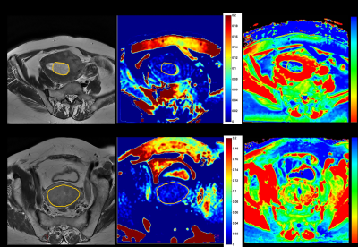 |
Evaluation of Amide Proton Transfer-Weighted Imaging and T2
mapping for preoperative risk stratification of endometrioid
adenocarcinoma
Fang Wang1,
Lianhua Cheng1,
Lingyu Chang1,
Dmytro Pylypenko2,
Weiqiang Dou2,
Dexin Yu1,
and Qing Wang1
1Qilu Hospital of Shandong University, Jinan city, Shandong province, China, 2GE Healthcare, Beijing, China, Beijing, China Keywords: Uterus, Cancer, Amide proton transfer-weighted imaging; T2 mapping ; endometrioid adenocarcinoma; risk stratification Motivation: Preoperative risk stratification of endometrioid adenocarcinoma (EA) impacts the choice of operative modality and prognosis of patients. Goal(s): We aimed to perform an accurate and non-invasive preoperative risk stratification method for EA by MRI sequences. Approach: APTw imaging as well as T2 mapping were included in this study. Results: The APTw and T2 values showed significant differences between the low- and non-low-risk groups. Combining APTw with T2 mapping achieved the highest diagnostic efficacy in the preoperative risk stratification. Impact: This suggested that the integrated use of APTw and T2 mapping can be an effective method for the preoperative risk assessment of EA. |
| 13:45 |
0211.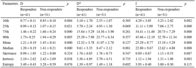 |
Pre-treatment multiparametric MRI for prediction of
chemo‑immunotherapy response in advanced non-small cell lung
cancer
Yu Zheng1 and
Jing Zhang1
1Lanzhou University Second Hospital, Lanzhou, China Keywords: Lung, Cancer Motivation: Early detection in poor responders to chemo‑immunotherapy for non-small cell lung cancer (NSCLC) facilitates timely adjustment of treatment strategies. Goal(s): To explore the value of histogram analysis based on intravoxel incoherent motion (IVIM) and difusion kurtosis imaging (DKI) in predicting chemo‑immunotherapy response in advanced NSCLC. Approach: 72 NSCLC patients underwent pre-treatment MRI examination. Histogram parameters of IVIM and DKI were calculated and compared. Results: Compared with non-responders, ADC, D, Dapp were significantly lower and f was higher in responders (all p <0.05). The multivariate logistic regression model performed the best with an AUC of 0.954. Impact: IVIM and DKI imaging would predict chemo-immunotherapy response in advanced NSCLC at initial state, which could help make clinical individualized treatment strategies. |
 Back to Meeting Home
 Back to the Program-at-a-Glance
The International Society for Magnetic Resonance in Medicine is accredited by the Accreditation Council for Continuing Medical Education to provide continuing medical education for physicians.
 View Presentation Video