Traditional Poster
Quantitative Imaging for Body Applications
ISMRM & ISMRT Annual Meeting & Exhibition • 04-09 May 2024 • Singapore

Traditional Poster
Quantitative Imaging for Body Applications
4815.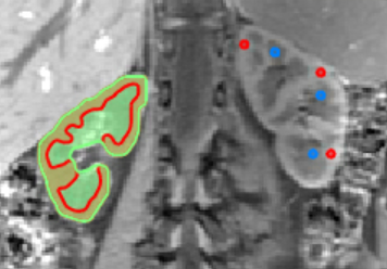 |
Test-retest repeatability of renal MRI parameters in healthy
volunteers comparing ROI-based and tissue segmentation based
image analysis
Cecilia Liang1,
Isabelle Loster2,
Thomas Küstner1,3,
Brigitte Gückel1,
Bernd Kühn4,
Fritz Schick3,
Ferdinand Seith1,
and Petros Martirosian3
1Department of Diagnostic and Interventional Radiology, University Hospital Tuebingen, Tuebingen, Germany, 2University Tuebingen, Tuebingen, Germany, 3Section on Experimental Radiology, University Hospital Tuebingen, Tuebingen, Germany, 4Siemens Healthineers, Erlangen, Germany Keywords: Kidney, Kidney Motivation: Multiparametric MRI of the kidneys is a promising technique for renal diagnostics, but the diversity of imaging protocols and analysis strategies hinders clinical translation. Goal(s): Our goal was to assess the repeatability of a multiparametric MRI protocol comparing ROI-based and tissue segmentation based analysis. Approach: Ten volunteers were examined with a multiparametric MRI protocol including ASL, IVIM, BOLD, T1 and T2 mapping twice with one week between visits. Results: Good repeatability of the multiparametric protocol could be achieved. T1 and T2 values showed less variability compared to perfusion and diffusion related functional parameters. Tissue segmentation showed better repeatability compared to ROI-based analysis. Impact: Our study demonstrates relatively high repeatability of multiparametric functional MRI of the kidneys. The results of the image analysis methods suggest, that manual segmentation is to be preferred over ROI-based analysis, if automated segmentation is not available. |
|
4816.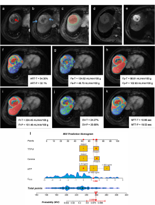 |
Bi-regional quantitative DCE-MRI for prediction of microvascular
invasion in hepatocellular carcinoma and its significance for
treatment
Yongjian Zhu1,
Peng Wang1,
Wei Cai1,
Bingzhi Wang2,
Xuan Meng3,
Sicong Wang4,
and Xiaohong Ma1
1Department of Diagnostic Radiology, National Cancer Center/National Clinical Research Center for Cancer/Cancer Hospital, Chinese Academy of Medical Sciences and Peking Union Medical College, Beijing, China, 2Department of Pathology, National Cancer Center/National Clinical Research Center for Cancer/Cancer Hospital, Chinese Academy of Medical Sciences and Peking Union Medical College, Beijing, China, 3Department of Hepatobiliary Surgery, National Cancer Center/National Clinical Research Center for Cancer/Cancer Hospital, Chinese Academy of Medical Sciences and Peking Union Medical College, Beijing, China, 4GE Healthcare, MR Research China, Beijing, China Keywords: Liver, Liver Motivation: Accurately predicting microvascular invasion (MVI) risk in hepatocellular carcinoma before surgery could aid clinicians in selecting appropriate surgical approaches to improve the patient’s prognosis. Goal(s): To construct DCE-MRI based nomogram for predicting MVI, and to assess its ability for stratifying the risk of recurrence after hepatectomy and guiding surgical approaches. Approach: Quantitative DCE-MRI parameters from both intra-tumoral region (ITR) and peritumoral region (PTR), along with clinical-radiological (CR) features, were utilized to establish the nomogram. Results: The nomogram presented AUC values of 0.966 in the training and 0.937 in the validation set for predicting MVI. High-risk patients could obtain survival benefit from anatomical resection. Impact: We constructed and evaluated the performance of the bi-regional quantitative DCE-MRI based nomogram for predicting MVI risk in HCC. Our predictive model effectively predicts MVI risk and assists clinicians in selecting appropriate therapeutic strategies for patients. |
|
4817.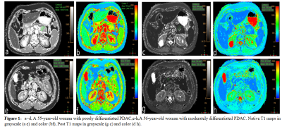 |
The value of Native T1 mapping in identifying pancreatic ductal
adenocarcinomas with different degree of pathological
differentiation
Kai Li1,
HaiYan Chen1,
Lei Shi1,
Rong Li Tang1,
and Shan Huang2
1Zhejiang Cancer Hospital, Hangzhou City,Zhejiang Province, China, 2Philips Healthcare, Shanghai, China Keywords: Cancer, Relaxometry, T1-mapping Motivation: MRI quantitative technique T1 mapping can reflect the change of intrinsic information of tissues Goal(s): To explore the value of T1 mapping in PDCA with different degrees of differentiation Approach: Comparison of PDCA with different degrees of differentiation, the T1 relaxation time before and after enhancement Results: by comparing the T1 mapping values before and after enhancement of PDAC with different degrees of differentiation, it was found that as the degree of differentiation of PDAC was getting higher, the Native T1 value showed a tendency to increase. The Native T1 was moderately positively correlated with the degree of pathological differentiation Impact: T1 mapping may serve as a noninvasive means of predicting the degree of pathologic differentiation of PDAC before surgery.Give clinicians earlier information. |
|
4818.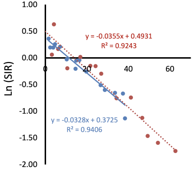 |
Field and TE independent liver iron concentration estimation
using signal intensity ratios
Eamon C Doyle1 and
John C Wood2
1Radiology, Children's Hospital of Los Angeles-USC KSOM, Los Angeles, CA, United States, 2Pediatrics and Radiology, Children's Hospital of Los Angeles-USC KSOM, Los Angeles, CA, United States Keywords: Liver, Relaxometry, Iron Overload Motivation: Estimation of liver iron concentration by R2* relaxation (LICR2*) is a powerful and widely used technique, however, it may fail from signal loss at high liver iron concentration. Goal(s): To estimate LIC from a single-TE liver-muscle signal intensity ratio (LICSIR) and validate at 1.5 and 3.0 Tesla. Approach: Using LICR2* estimates collected at 1.5T as a reference, we compared LICSIR estimates in 15 subjects who had undergone MRI examination at both 1.5T and 3.0T. Results: We were able to derive field-independent scaling constants that allow LICSIR estimation at 1.5 and 3.0T, more than doubling the effective dynamic range of LICR2* estimation. Impact: This generalized framework for LICSIR estimation allows reasonable LIC values to be reported (and trended) in patients for whom traditional relaxometry has failed. It also allows approximate LIC calculation from commonly used single and dual echo gradient echo acquisitions. |
|
4819.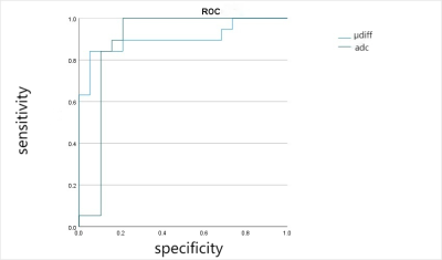 |
Value of placental stiffness using virtual magnetic resonance
elastography in pregnancies complicated by pre-eclampsia
Jialu Xu1 and
Jiejun Cheng1
1Department of Radiology, Shanghai First Maternity and Infant Hospital , Tongji University, Shanghai, China Keywords: Placenta, Placenta Motivation: To evaluate virtual magnetic resonance elastography in healthy and preeclamptic(PE) pregnancies. Goal(s): To compare the stiffness value (μdiff) and apparent diffusion coeffificient (ADC) in healthy and PE pregnancies. Approach: DWI(b-value of 50 ,200and 800 s/mm2 ) were performed on all pregnant women using a 1.5 T MRI scanner. The value of μdiff and ADC were calculated and compared between groups. Results: The mean ADC value of the control and PE groups were 1.414±0.228×10⁻³ mm2/s and 1.737±0.107×10⁻³ mm2/s, respectively. The mean μdiff value of PE were 5.901±1.757 and 4.618±2.055 kPa, respectively. The area under the curve for μdiff was 0.903 and ADC was 0.796, respectively. Impact: Placental μdiff value was found to be more reliable than ADC in differentiating between normal and preeclampsia placentas. |
|
4820.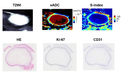 |
DWI Parameters Correlated with Ki-67 Expression, a Prognostic
Biomarker, in a Triple-Negative Breast Cancer PDX Mouse Model
Mami Iima1,2,
Marino Akamatsu3,
Hirohiko Imai4,
Hana Suzuki3,
Noriko Gotoh5,
Yasuto Takeuchi5,
Maya Honda1,
Tomomi Nobashi1,
Masako Kataoka1,
Akihiko Yoshizawa6,
hiroaki Ito7,
Tomoe Nakagawa8,
Minsoo Kim9,
Denis Le Bihan10,11,
and Yuji Nakamoto1
1Diagnostic Imaging and Nuclear Medicine, Kyoto University Graduate School of Medicine, Kyoto, Japan, 2Institute for Advancement of Clinical and Translational Science (iACT), Kyoto University Hospital, Kyoto, Japan, 3Faculty of Medicine, Kyoto University, Kyoto, Japan, 4Department of Systems science, Graduate School of Informatics, Kyoto University, Kyoto, Japan, 5Cancer Research Institute, Kanazawa University, Kanazawa, Japan, 6Diagnostic Pathology, Nara Medical University, Nara, Japan, 7Diagnostic Pathology, Kyoto University Graduate School of Medicine, Kyoto, Japan, 8Department of Pathology and Tumor Biology, Kyoto University Graduate School of Medicine, Kyoto, Japan, 9Laboratory of Integrative Molecular Medicine, Kyoto University Graduate School of Medicine, Kyoto, Japan, 10Human Brain Research Center, Kyoto University Graduate School of Medicine, Kyoto, Japan, 11CEA-Saclay, Paris-Saclay University, NeuroSpin, Gif/Yvette, France Keywords: Cancer, Breast Motivation: The lack of reliable biomarkers for assessing tumor characteristics and the limitations of histological analysis due to tumor heterogeneity led to the exploration of diffusion-weighted parameters. Goal(s): To investigate the association between DW parameters and Ki-67 expression in triple-negative breast cancer, with a focus on whether they can serve as prognostic biomarkers. Approach: Seventeen triple-negative breast cancer mice with a PDX model underwent 7T MRI scans, yielding DW images. Advanced analysis evaluated ADC and non-Gaussian diffusion parameters, validated through histological Ki-67 staining. Results: DWI parameters (S-index, sADC, and ADC0) show strong correlations with Ki67 levels, at short and long diffusion times (9, 27.6ms). Impact: Promising prognostic biomarkers for triple-negative breast cancer, DWI parameters (S-index, sADC, and ADC0) displayed strong correlations with Ki-67 expression, at short and long diffusion times. This validation through accurate DWI-pathology comparison highlights imaging's pivotal role in advancing breast cancer diagnosis. |
|
4821. |
The value of IVIM-DWI and DCE-MRI in predicting molecular
subtypes of breast cancer
Ting-ting Lin1
1The First Affiliated Hospital of USTC(Anhui Provincial Cancer Hospital), Hefei, China Keywords: Breast, Cancer, molecular subtypes Motivation: To analyze the value of imaging examination in accurate diagnosis of breast cancer. Goal(s): Provide an non-invasive examination for the prediction of molecular subtypes of breast cancer before treatment. Approach: The quantitative parameters of IVIM-DWI and DCE-MRI in patients with breast cancer without any invasive examination and treatment were compared with pathological molecular subtypes, so as to analyze their diagnostic value in the prediction of molecular subtypes. Results: DCE-MRI and IVIM-DWI were correlated with immune prognostic factors of breast cancer and had differential diagnostic value for different molecular subtypes. Impact: DCE-MRI and IVIM-DWI could provide a non-invasive diagnostic method for predicting molecular subtypes of breast cancer and provide a reference for further development of personalized treatment plans. |
|
4822.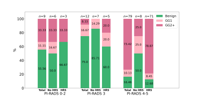 |
Quantitative Imaging Habitat Risk Score (HRS) Combined with
PI-RADSv2 Improves Predictive Value of Prostate Lesion
Identification on mpMRI
Veronica Wallaengen1,
Adrian L Breto1,
Isabella M Kimbel1,
Evangelia I Zacharaki1,
Ahmad Algohary1,
Nachiketh Soodana Prakash2,
Sandra M Gaston1,
Rosa P Castillo Acosta3,
Oleksandr N Kryvenko4,
Bruno Nahar2,
Dipen J Parekh2,
Alan Pollack1,
Sanoj Punnen2,
and Radka Stoyanova1
1Department of Radiation Oncology, University of Miami Miller School of Medicine, Miami, FL, United States, 2Department of Urology, University of Miami Miller School of Medicine, Miami, FL, United States, 3Department of Radiology, University of Miami Miller School of Medicine, Miami, FL, United States, 4Department of Pathology, University of Miami Miller School of Medicine, Miami, FL, United States Keywords: Software Tools, Cancer Motivation: Discrimination between true and false positive targets identified using PI-RADSv2 is needed to avoid unnecessary biopsies. Goal(s): To investigate how quantitative analysis of prostate mpMRI through Habitat Risk Scoring (HRS) combined with PI-RADS can improve prostate lesion identification compared to using PI-RADS alone. Approach: In prospective clinical trials lesions identified by PI-RADS and/or HRS were targeted through MRI/ultrasound fusion biopsies. Results: Using HRS yields 100% NPV in the PI-RADS 3 cohort and increases PPV by 7.4% in the PI-RADS 4-5 cohort with respect to clinically significant cancer. Overall, the NPV and PPV increased by 21.4% and 13.2% respectively. Impact: We present a quantitative imaging approach to complement the current standard for assessing prostate cancer risk in mpMRI data and demonstrate that the use of HRS strengthens fidelity in both positive and negative detections. |
|
4823.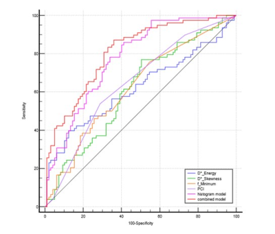 |
Intravoxel incoherent motion diffusion-weighted imaging in
diagnosing perineural invasion status of rectal cancer: a
histogram analysis study
Rong He1,
Gesheng Song1,
Junyi Fu1,
Weiqiang Dou2,
Aiyin Li1,
and Jingbo Chen3
1Department of Radiology, The First Affiliated Hospital of Shandong First Medical University & Shandong Provincial Qianfoshan Hospital, Jinan, China, 2GE Healthcare,MR Research, Beijing, China, 3Department of General Surgery, The First Affiliated Hospital of Shandong First Medical University & Shandong Provincial Qianfoshan Hospital, Jinan, China Keywords: fMRI Analysis, Diffusion/other diffusion imaging techniques, histogram analysis Motivation: Currently, perineural invasion (PNI) of rectal cancer (RC) can only be confirmed by pathological examination of postoperative specimens. Goal(s): It aimed to investigate the intravoxel incoherent motion (IVIM) diffusion-weighted imaging (DWI) in diagnosing the PNI status of RC by histogram analysis.
Approach: We extracted histogram features from 7
parametric maps derived from IVIM-DWI. The independent
predictive histogram features of rectal cancer PNI were
combined with the percentage of rectal wall Results: The AUC of the combined model is higher than that of each single-parameter model and histogram model. Impact: This study demonstrated that full-volume histogram parameters basing on IVIM-DWI can be used to assess PNI status in rectal cancer. Histogram analysis, as a non-invasive tool, may be more valuable not only in rectal cancer research in the future. |
|
4824.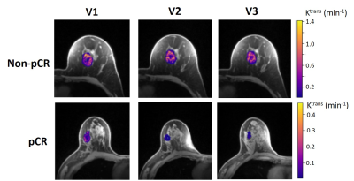 |
Multi-Center and Multi-Vendor Platform DCE-MRI Prediction of
Breast Cancer Therapy Response: A Preliminary Comparison of
Imaging Biomarkers
Brendan Moloney1,
Xin Li1,
Michael Hirano2,
Assim Saad Eddin3,
Jeong Youn Lim1,
Debosmita Biswas2,
Anum S. Kazerouni2,
Alina Tudorica1,
Isabella Li2,
Mary Lynn Bryant2,
Courtney Wille3,
Chelsea Pyle1,
Habib Rahbar2,
Su Kim Hsieh3,
Travis Rice-Stitt1,
Suzanne Dintzis2,
Amani Bashir3,
Evthokia Hobbs1,
Alexandra Zimmer1,
Jennifer Specht2,
Sneha Phadke3,
Nicole Fleege3,
James Holmes3,
Savannah C. Partridge2,
and Wei Huang1
1Oregon Health & Science University, Portland, OR, United States, 2University of Washington, Seattle, WA, United States, 3University of Iowa, Iowa City, IA, United States Keywords: Treatment Response, Cancer, Multi-Center and Multi-Vendor platform, DCE-MRI, Therapy Response, Ktrans Motivation: Validate Shutter-Speed model (SSM) DCE-MRI as a robust predictor of breast cancer (BC) response to neoadjuvant chemotherapy (NAC) in a multi-center and multi-vendor platform setting. Goal(s): Compare tumor size, semi-quantitative, and quantitative DCE-MRI for early prediction of NAC response. Approach: BC patients treated with NAC underwent longitudinal high spatiotemporal resolution DCE-MRI at three sites using a 3T Siemens, GE, or Philips system. Semi-quantitative signal-enhancement-ratio (SER) and quantitative Tofts model (TM) and SSM pharmacokinetic (PK) parameters were derived from DCE time-course data. Results: PK parameters outperformed size and SER while SSM was superior to TM in early prediction of pathologic response. Impact: It is feasible to implement quantitative high spatiotemporal resolution SSM DCE-MRI in trials with multi-center and multi-vendor platform settings for robust assessment of BC response to NAC. |
|
4825.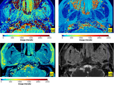 |
Synthetic MRI for the quantitative Assessment of efficacy of
Immunotherapy in nasopharyngeal carcinoma.
Yu Huang1 and
Chen ZHAO2
1Radiology, The First Clinical Medical College of Guangxi Medical University, Nanning, China, 2MR Research Collaboration, Siemens Healthineers., Nanning, China Keywords: Other Interventional, Quantitative Imaging Motivation: Due to the internal heterogeneity of the nasopharyngeal carcinoma, conventional treatment fails to enhance the tumor microenvironment, ultimately leading to recurrence or metastasis. Goal(s): To investigate whether quantitative values derived from T1, T2, and PD maps can serve as an assessment index for combined immunotherapy with chemotherapy in patients with newly diagnosed locally advanced NPC. Approach: Synthetic MRI, non-invasive technique, can generate quantitative values for intrinsic tissue features. Results: The study indicate the potential of PD and T2 values in distinguishing between two treatment. A larger sample size is required to further validate the value of SyMRI in evaluating immunotherapy for NPC. Impact: Synthetic MRI can generate longitudinal relaxation time (T1), transverse relaxation time (T2), and proton density (PD). |
|
 Back to Meeting Home
 Back to the Program-at-a-Glance
The International Society for Magnetic Resonance in Medicine is accredited by the Accreditation Council for Continuing Medical Education to provide continuing medical education for physicians.