Weekend Course
X-Nuclei & Spectroscopy: Everything, Everywhere but Not Quite All at Once
ISMRM & ISMRT Annual Meeting & Exhibition • 04-09 May 2024 • Singapore

Weekend Course
X-Nuclei & Spectroscopy: Everything, Everywhere but Not Quite All at Once
| 08:00 |
Clinical Applications of MRS & MRSI in the Brain
Evita Wiegers
Keywords: Contrast mechanisms: Spectroscopy Magnetic Resonance Spectroscopy (MRS) provides a unique window into tissue composition and metabolism. Its integration into clinical practice has paved the way for a multitude of applications. This educational session delves into the diverse clinical landscape where MRS plays a role, including the brain tumors, neurodegenerative disorders, and neonatal encephalopathies |
|
| 08:25 |
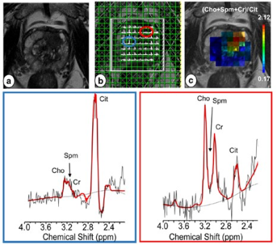 |
Clinical Applications of MRS in the Body
Saadallah Ramadan, Oun Al-iedani
Keywords: Body: Body, Contrast mechanisms: Spectroscopy, Education Committee: Clinical MRI Motivation:MR Spectroscopy (MRS) offers a unique perspective into the body's biochemistry, providing insights into various diseases beyond what conventional imaging techniques can reveal.Goal(s):To elucidate the clinical applications of MRS, highlighting its role in diagnosing and monitoring diseases. Approach:To explore MRS techniques, examining its integration with MRI, and discussing specific applications in various pathologies through case studies and recent research. Results:MRS's effectiveness in precise diagnosis and treatment monitoring,revealing its potential in clinical scenarios from cancer to metabolic disorders. Impact:MRS's capabilities are highlighted, prompting further research into its diagnostic precision. This enables clinicians to transform how they care for their patients. |
| 08:50 |
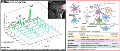 |
Diffusion-Weighted MRS: Insights into the Brain
Microstructure from Metabolites
André Döring
Keywords: Contrast mechanisms: Diffusion-Weighted MR Spectroscopy, Contrast mechanisms: Diffusion Magnetic resonance spectroscopy (MRS) allows quantifying concentrations of cell-type specific metabolites. This enabled to monitor metabolic pathways. However, alongside metabolic changes, alterations in cellular microstructure represent pivotal pathomechanistic shifts in the onset and progression of neurodegenerative-diseases (e.g., glial morphology in neuroinflammation or neuronal integrity in cerebral atrophy). By integrating diffusion-weighting into MRS (dMRS), we can measure the diffusion properties of individual, celltype-specific metabolites that can serve as probes for cellular microstructure. In combination with simulation techniques, enabling an absolute quantification of cellular-features, dMRS holds immense promise in offering a new class of biomarkers for identifying cell-type specific (patho)morphological alterations. |
| 09:15 |
Functional MRS: Neurotransmitters & Brain Metabolism
Francesca Branzoli
Keywords: Contrast mechanisms: Spectroscopy, Neuro: Brain function Functional MRS (fMRS) aims to measure, noninvasively, subtle changes in metabolic concentrations upon neural activation induced by sensory or cognitive tasks. fMRS may provide insights onto energy metabolism and brain function that are not captured by functional MRI, which is based on the blood-oxygenation level dependent effect. In this presentation, I will provide an overview of the metabolic changes that have been detected during neuronal activity and will discuss how these findings contributed to our knowledge of brain energy metabolism and neurotransmission. FMRS technical challenges and limitations will also be reviewed. |
|
| 09:40 |
Break & Meet the Teachers |
|
| 10:10 |
CEST: Covering the Full Spectrum
Nirbhay Yadav
Keywords: Contrast mechanisms: CEST & MT, Contrast mechanisms: Molecular Imaging, Physics & Engineering: Physics Chemical Exchange Saturation Transfer (CEST) signals are often not visible in the MRS spectrum due to exchange-dependent line broadening and/or the effects of water presaturation transferring to solute signals. Conversely, CEST relies on the presaturation of solute signals transferring to water and then detecting the accumulated partial saturation of water. This process retains the molecular specificity of spectroscopy techniques and, the repeated label-tranfer effect during the presaturation period, results in a sensitivity enhancement that is orders of magnitude beyond the solute molecular concentration. This presentation will describe several different types of CEST signals and factors that determine their amplitude. |
|
| 10:35 |
X-Nuclei, biomolecules and their application
Yoichi Takakusagi
Keywords: Contrast mechanisms: Non-proton, Contrast mechanisms: Spectroscopy, Contrast mechanisms: Hyperpolarized MR (Non-Gas) Nuclear magnetic resonance (NMR) detects the nuclear spin behavior of observable nuclides as spectra or images using radio frequency (RF) pulses. The H nucleus, which is a component in water molecules that make up 65-70% of the human body, is the main nuclide responsible for MR image acquisition. On the other hand, there are some X-nuclei, which can also be observed via NMR and possess distinctive characteristics unlike H. In this presentation, representative X-nuclei and the biomolecules containing the nuclei will be introduced, associating with the relationship with commonly used MRI elemental techniques. |
|
| 11:00 |
A Neutron & a Proton: 101 Guide to Deuterium Imaging
Qingjia Bao
|
|
| 11:25 |
 |
Advanced Methods in Deuterium Imaging
Fabian Niess
Keywords: Contrast mechanisms: Deuterium Deuterium metabolic imaging is an emerging magnetic resonance technique for noninvasive imaging of glucose metabolism. Short relaxation times allows 3D imaging with reasonable spatial resolution in clinical feasible scan times using conventional phase encoding approaches. Over the last few years this method has been used extensively by the majority of scientific sites from ultra-high to clinical magnetic field strengths. Recently, novel approaches have been presented to simultaneously obtain complementary anatomical data, boost the achievable SNR and accelerate data acquisition to achieve higher spatial resolution without increasing scan times. This presentation summarises recent developments of advanced methods in deuterium metabolic imaging. |
 Back to Meeting Home
 Back to the Program-at-a-Glance
The International Society for Magnetic Resonance in Medicine is accredited by the Accreditation Council for Continuing Medical Education to provide continuing medical education for physicians.
 View Presentation Video