Weekend Course
Gender Imaging: Prostate & Female Pelvis
ISMRM & ISMRT Annual Meeting & Exhibition • 04-09 May 2024 • Singapore

Weekend Course
Gender Imaging: Prostate & Female Pelvis
| 13:00 |
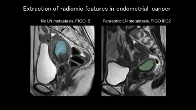 |
Uterine Cancer: T2W or Texture?
Yuki Himoto
Keywords: Body: Urogenital Motivation: Conventional MRI is crucial for uterine cancer in clinical settings. Radiomics, utilizing quantitative imaging features, has rapidly evolved in research. A brief review provides insights for addressing current clinical challenges and guiding future developments. Goal(s): The goals are to grasp the roles of conventional MRI, achievements in radiomic studies for uterine cancer (particularly endometrial cancer), and the limitations and improvements in imaging quality. Approach: Reviewing the latest research on MRI and radiomics in uterine cancer, with a primary focus on endometrial cancer. Results: Despite limitations, radiomics continues to advance. Improvements in conventional MRI quality for gynecologic imaging are clinically significant and enhance radiomics. Impact: Grasping current improvements in MRI quality and radiomics, along with identifying challenges, offers insights for addressing clinically relevant issues in uterine cancer. |
| 13:25 |
Cervical Cancer: T2W or Texture?
Luca Russo
Keywords: Body: Urogenital
|
|
| 13:50 |
Ovarian Cancer: T2W or Texture?
Aki Kido
Keywords: Body: Pelvis Motivation:T2WI, i.e. traditional morphological image diagnosis is good at ‘present’ histopathological diagnosis. On the other hand, recent texture, i.e. radiomics analysis can provide invisible information and make ‘future’ diagnosis including prognosis and treatment response.Goal(s): Clinical radiologists need to know the pros and cons of both morphological diagnosis and texture analysis and know how to get the necessary information when we need it. Approach: - Results: - Impact: - |
|
| 14:15 |
Advanced Morphofunctional Sequences & AI in the Evaluation of
Fetal Growth Anomalies
Lucia Manganaro
Keywords: Image acquisition: Image processing, Image acquisition: Sequences, Image acquisition: Motion Correction Intrauterine growth restriction (IUGR) is the fetal failure to reach its biological growth potential and placental dysfunction is the main cause IUGR is distinguished into Small for Gestational Age (SGA) and Fetal Growth Restriction (FGR). Ultrasound represents the technique of choice for the diagnosis and the Functional Doppler parameters improve this distinction as established by the Expert Consensus Statement. The differentiation is important: many SGAs are constitutionally small but healthy without risk of poor outcomes unlike FGRs.Magnetic Resonance Imaging (MRI) and Diffusion weighted imaging (DWI) can evaluate placenta and fetal growth supporting the diagnosis of developmental pathologies |
|
| 14:40 |
Break & Meet the Teachers |
|
| 15:10 |
 |
The Problem with PI-RADS
Yan Mee Law
Keywords: Body: Urogenital, Cross-organ: Cancer, Image acquisition: Multiparametric PI-RADS v2.1 is widely adopted by the international radiology and urology communities in risk assessment of clinically significant prostate cancer (csPCa) at multi-parametric MRI (mpMRI) in treatment naïve men. This consensus document, aimed at establishing uniformity in acquisition and interpretation of mpMRI, has been prone to performance variations. A non-negligible number of csPCa are missed at mpMRI as these lesions resemble benign lesions or possess mpMRI features that do not fit a defined PIRADS category. We discuss the limitations and controversies of PIRADS and tips and tricks that may be useful to improve the conspicuity of “MRI occult” csPCa. |
| 15:35 |
Radiomic & Radiogenomic Signatures in Prostate Cancer
Daniel Moses
Keywords: Body: Pelvis, Body: Urogenital, Education Committee: Clinical MRI Radiomics and genomics have potential to add value in the diagnosis and risk stratification of cancer. Radiomics analyses quantitative imaging biomarkers of tumours in medical images, such as morphological, histogram and texture features, whereas genomics investigates genetic elements of cancer tissue. Combining these into radiogenomics enables the generation of combinations of features, known as signatures, that can predict cancer phenotypes which effect the biological properties and therefore the behaviour of tumours. This presentation examines MRI radiomics and radiogenomics and their use in aiding the screening, detection, classification, treatment, and prognosis of prostate cancer. |
|
| 16:00 |
Two “B” or Not Two “B”: Probing Prostate Tissue Composition
Aritrick Chatterjee
Keywords: Body: Pelvis, Contrast mechanisms: Microstructure, Image acquisition: Quantification Diffusion MRI is a key component of prostate MRI. A minimum of 2 b-values are needed to derive ADC map. PIRADS guidelines recommend a minimum of 3 b-values: low (0-100 sec/mm2), intermediate (800-1000 sec/m2) and high b-value (≥1400 sec/m2, either acquired or calculated). The talk will highlight how the choice of b-values can significantly affect ADC estimates.Additionally, we will look at recent advancements in quantitative MRI techniques that probe prostate tissue microstructure. We will look at imaging parameters used and results from these techniques: VERDICT, Restriction spectrum imaging, Hybrid Multi-dimensional MRI, time-dependent diffusion imaging, diffusion-relaxation correlation spectrum imaging, etc. |
|
| 16:25 |
 |
Exploiting AI in Prostate Cancer Assessment
Amit Mehndiratta
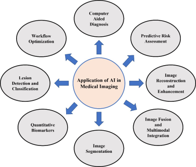
Keywords: Body: Pelvis, Cross-organ: Cancer, Image acquisition: Machine learning Prostate cancer is sixth leading cause of cancer related death and is one of the most prevalent malignancies in men. Artificial intelligence (AI) methods are potentially useful in prostate cancer management for detection and characterization of prostate lesion. AI solutions are assisting in streamlining patient workflow and optimizing treatment plans for individual patient leading to precision medicine-based approach. Machine learning methods are being used for segmentation of prostate gland and its anatomical structures, image registration, detection of lesions, lesion characterization, automated PI-RADS scoring, and risk stratification, which ultimately leads to enhanced diagnosis, treatment planning, and patient outcomes in prostate-related conditions. |
 Back to Meeting Home
 Back to the Program-at-a-Glance
The International Society for Magnetic Resonance in Medicine is accredited by the Accreditation Council for Continuing Medical Education to provide continuing medical education for physicians.
 View Presentation Video