Weekend Course
Basics of Cardiovascular MR
ISMRM & ISMRT Annual Meeting & Exhibition • 04-09 May 2024 • Singapore

| 07:45 |
Segmented & Rapid Imaging: Methods
Jennifer Steeden
Keywords: Image acquisition: Fast imaging, Cardiovascular: Cardiac, Image acquisition: Sequences MRI is an extremely valuable imaging technique, but it is inherently slow. This results in long acquisition times and sensitivity to motion. This makes imaging of the heart particularly challenging due to both cardiac and respiratory motion. There has been a lot of work on speeding up acquisition of data – many of these rely on efficient trajectories, as well as reducing the amount of data collected (including undersampling). This presentation will cover the main methods for speeding up cardiac MRI and the main advantages and disadvantages of each method. |
|
| 08:10 |
Segmented & Rapid Imaging: Applications
Teo Lynette
Keywords: Image acquisition: Fast imaging, Cardiovascular: Cardiac function, Cardiovascular: Myocardium Time is of the essence in any busy MRI service and CMR scans take up a significant proportion of scanner time. We have the tools for rapid image acquisition but many of us find it difficult to break away from segmented imaging techniques as accelerated acquisitions often come at the expense of image quality. There are concerns that subtle abnormalities in wall motion or small amounts of myocardial fibrosis may be missed using such techniques. This talk will illustrate segmented and rapid imaging techniques and it is for the user to decide what is best for their constraints and practice. |
|
| 08:35 |
Tissue Characterization in CMR: Methods
Masa Bozic-Iven
Cardiac magnetic resonance imaging (CMR) offers a plethora
of contrast mechanisms to assess cardiac health and plays a
major role in the management of cardiovascular diseases.
Beyond basic anatomical imaging, CMR provides tools for a
comprehensive evaluation of cardiac function. These range
from cine imaging to parametric mapping for quantitative
viability assessment. This presentation will briefly
introduce the basic physics behind common CMR sequences and
established methods, such as T1 and T2 mapping. Moreover, we
will explore advanced techniques, like late gadolinium
enhancement and myocardial perfusion imaging, as well as
novel contrast mechanisms including T1rho and diffusion
tensor imaging.
|
|
| 09:00 |
Tissue Characterization in CMR: Applications
Jeanette Schulz-Menger
|
|
| 09:25 |
Break & Meet the Teachers |
|
| 09:50 |
Myocardial Perfusion Imaging: Methods
Behzad Sharif
Keywords: Cardiovascular: Myocardium, Cardiovascular: Blood Cardiovascular MRI is a comprehensive modality for assessment of ischemic heart disease. Myocardial perfusion MRI captures the rapid dynamics of the first pass of a T1-shortening contrast agent through the coronary microvascular network. Vasodilator or exercise stress myocardial perfusion MRI is a powerful modality for diagnosis of ischemic heart disease (obstructive coronary artery disease and/or coronary microvascular dysfunction). We will review the basics and will then discuss several methodological advances introduced in the past decade. |
|
| 10:15 |
 |
Myocardial Perfusion Imaging: Applications
Stephen Cheung
Keywords: Cardiovascular: Cardiac, Cardiovascular: Hemodynamics, Cardiovascular: Myocardium Quantitative myocardial perfusion is now ready for clinical use and is available on platforms provided for vendors. It adds to qualitative perfusion by providing additional information about regional myocardial blood flow and reserve. After excluding artefacts the perfusion maps provide valuable overall impression of perfusion status and a normal map is a reliable exclusion of significant ischemia. Quantitative perfusion also enable diagnosis of microvascular dysfunction which is not always possible with qualitative analysis. Diagnosis of triple vessel disease is also more confident since it does not depends on comparison with regions that are supposed to be of normal perfusion. |
| 10:40 |
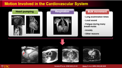 |
Motion in Cardiovascular MR: How the Heart Moves with the
Cardiac & Respiratory Cycles
Zhaoyang Fan
Keywords: Cardiovascular: Cardiovascular, Cardiovascular: Cardiac MR is a powerful imaging modality allowing for comprehensive cardiac imaging assessment. However, MR is highly motion sensitive and the cardiovascular system does involve several categories of motion, particularly respiratory-induced motion and cardiac pumping-induced motion, which will result in severe image degradation if not compensated. This talk will focus on introduction of these motion followed by overview of technical solutions to tackling them during cardiac MRI. |
| 11:05 |
Putting It All Together: An Efficient CMR Exam
Wendy Strugnell
Keywords: Cardiovascular: Cardiac As new imaging techniques are developed and the clinical applications of CMR expand, implementing efficient workflow practices has become increasingly important in clinical practice. To complete a comprehensive examination in a clinically acceptable timeframe with high quality imaging requires considerable forethought and planning. Developing and applying a systematic approach to all aspects of the examination can save considerable scanner time, even if the operator is proficient in the placement of imaging planes.This presentation will outline practical ways to improve the efficiency of a CMR exam in the clinical environment. |
 Back to Meeting Home
 Back to the Program-at-a-Glance
The International Society for Magnetic Resonance in Medicine is accredited by the Accreditation Council for Continuing Medical Education to provide continuing medical education for physicians.
 View Presentation Video