Weekend Course
Advanced Cardiovascular MRI Techniques
ISMRM & ISMRT Annual Meeting & Exhibition • 04-09 May 2024 • Singapore

Weekend Course
Advanced Cardiovascular MRI Techniques
| 13:15 |
Regional Function & Strain
Frederick Epstein
|
|
| 13:40 |
Real-Time & Free-Breathing Techniques
Haikun Qi
Keywords: Cardiovascular: Cardiac function, Image acquisition: Fast imaging, Image acquisition: Reconstruction Motivation: Real-time MRI can resolve cardiac and respiratory motion and has unique advantages for cardiac imaging. An overview of this active research area is necessary. Goal(s): Introduce various reconstruction methods of accelerated real-time imaging and highlight its applications. Approach: Undersampled real-time MRI reconstruction techniques improving the spatial-temporal resolution are covered, including k-t methods, parallel imaging, compressed sensing and low-rank methods, and recent advances that enable extreme acceleration. Results: Real-imaging imaging has demonstrated superior performance in cardiac function evaluation, flow analysis, and tissue characterization, and MR-guided treatment. Impact: The audience can grasp basic and advanced reconstruction techniques of real-time MRI and its potential applications. |
|
| 14:05 |
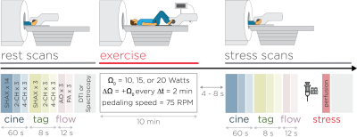 |
Cardiac MRI During Exercise
Manuel Morales
Keywords: Cardiovascular: Cardiovascular This educational talk presents an overview of cardiac MRI during exercise, highlighting its challenges and recent advancements. We explore technical specifications for optimal imaging during exercise and advancements in Ex-CMR sequences and image reconstruction techniques. The aim is to shed light on the complexities of capturing accurate cardiac images during physical stress and the potential of these techniques in diagnosing and managing cardiovascular diseases. |
| 14:30 |
Multiparametric & Fingerprinting in Cardiac MRI: Everything All
at Once
Claudia Prieto
|
|
| 14:55 |
Break & Meet the Teachers |
|
| 15:25 |
Cardiac MRI to Characterize Diastolic Function
Masaki Ishida
Keywords: Cardiovascular: Cardiac, Cardiovascular: Cardiac function, Cardiovascular: Myocardium Diastolic dysfunction, a significant contributor to HFpEF, presents a formidable diagnostic challenge due to its diverse etiologies and complex pathophysiology. While cardiac catheterization is currently considered the reference standard for assessing LV diastolic function, it has inherent limitations, notably its invasiveness. Cardiac MRI offers a range of non-invasive, objective, and reproducible measures of diastolic function, including ventricular and atrial volumes, myocardial strain, native T1 and ECV, 4D flow, and exercise real-time cine index. These advanced CMR techniques substantially enhance noninvasive and objective evaluation of diastolic function and refine clinical management strategies for patients with diastolic dysfunction and HFpEF. |
|
| 15:50 |
AI-Driven CMR
Chen Qin
Keywords: Image acquisition: Machine learning, Cardiovascular: Cardiac Artificial intelligence (AI) is making a significant impact on all aspects of cardiovascular magnetic resonance (CMR) imaging. In this talk, we will focus on discussing the recent development of DL in CMR imaging workflow, from the reconstruction of accelerated signals to automatic quantification of clinically useful information. Specifically, we will describe how DL methods can be used for reconstruction of accelerated dynamic cine CMR imaging. We will also show the utility of DL for CMR analysis, with a particular focus on CMR segmentation and motion tracking. Finally, we will briefly discuss about their current limitations, challenges, and future opportunities. |
|
| 16:15 |
Radiomics in CMR
Reza Nezafat
|
|
| 16:40 |
 |
4D Flow MRI
Susanne Schnell
Keywords: Cardiovascular: Hemodynamics, Contrast mechanisms: Flow, Image acquisition: Quantification Phase-contrast MRI is an angiography technique that uses bipolar gradients to encode tissue or fluid motion into the MRI signal phase. If measured in 2D with 1-directional encoding this is termed 2D Phase-Contrast MRI. Extended to a time-resolved heart or pulse rate-triggered 3D sequence with 3-directional velocity encoding, this is termed 4D flow MRI. Error sources will be described throughout the course as well as data analysis and quantification approaches. Further extensions such as dual- and multi-venc, and 5D flow MRI will be described. Finally, applications in the thorax, head, and abdomen will be introduced. |
 Back to Meeting Home
 Back to the Program-at-a-Glance
The International Society for Magnetic Resonance in Medicine is accredited by the Accreditation Council for Continuing Medical Education to provide continuing medical education for physicians.
 View Presentation Video