Wednesday 26 April
Jump to:
Don’t Miss Today:
Plenary Session: Nanotheranostics is a rapidly growing field at the interface of diagnosis and therapy. In this Plenary session, Dr. Hadjipanayis will discuss the simultaneous targeting, treatment and imaging of malignant brain tumors with magnetic nanoparticles. Dr. Bhujwalla will describe the delivery of therapy to cancer cells, stromal cells and microenvironments while perserving the surrounding healthy tissue. Dr. Niendorf will make a case for routinely monitoring drug distribution in rheumatoid arthritis patients by using of fluorine (19F) MR. These technologies are providing exciting new roles for MRI and MRS in combined imaging and therapeutic strategies en route to personalized medicine.
Inaugural Junior Fellows Symposium: Machine Learning in Imaging (1:45-3:45pm, 313BC). As a new initiative the junior fellows organzied the entire program, which featuers talks by Daniel Alexander, Kerstin Hammernik and Dinggang Shen, followed by a panel discussion on clinical integration.
Special Session – Scientific Highlights: At the end of the plenary session, five top-scoring abstracts from the conference will be featured (#750-754). These include Dynamic Diffusion MRI Signal Changes Accompany Electrical Activity in Myelinated Axons, Separating positive and negative susceptibility sources in QSM, Spatiotemporal analysis of breathing-induced fields in the cervical spinal cord at 7T, Using Machine Learning to study knee Osteoarthritis: the path towards OA Precision Medicine, and Mapping of Abnormal Aortic Hemodynamics in 515 Patients with Aortopathy.

In the Resonarium: Process, challenges and pitfalls of successful algorithm development (9-10am), and BART Reconstruction demo (1:45-2:45pm), Machine Learning (5-6pm)
Techniques for personalized MRI: Education session 7-7:50am in 313BC features Drs. Duygu Tosun and Andreia Faria discussing design and application of single-subject and atlas-based brain analysis.
What’s Hot:
Sunrise Session – Gadolinium in MSK Imaging: The role of gadolinium enhancement in assessment of tumors and infection with emphasis on contrast kinetics including binding constants and kinetics of dissociation.
#0693 Fracture Risk using deep learning and hip microarchitecture MRI: Developed supervised convolutional neural networks for hip fracture risk identification using proximal femur MR microarchitecture images and patients’ history of fragility fractures.
Fool your surgeons! ZTE in the shoulder: (#0849) discusses the ability of ZTE to accurately assess osseous detail necessary for surgical planning in the shoulder using CT as the standard.
#0847 Feasibility of bone mineral and water quantification in vivo by solid–state 31P and H at 3T: This study suggests that both organic and inorganic phases of cortical bone can be quantitatively evaluated in vivo with a single, integrated protocol.
It Doesn’t Have to Be That Way: Information & Diagnosis, from 7:00-7:50 AM in Room 315. The talks will focus on novel compressed sensing approaches to removing artifacts and the subject that is seemingly on everyone’s mind – machine learning/artificial intelligence/computer assisted diagnosis

Another Step Towards Personalized Medicine: Stephanie Hectors and colleagues explore the use of multiple quantitative MR technologies to explore tumor heterogeneity. #661, Power Pitch Theater B, 08:15.
#675 – Simultaneous acquisition of MRA and ASL perfusion. Okell describes a novel approach for 4D acquisition that permits recovery of both whole-brain dynamic MRA and time-resolved PCASL ASL perfusion maps. (Arterial Spin Labeling: Making it More Robust & Informative, Room 310, 8:15 am)
#814 – Introducing TEdDI: Veraart and colleagues study the TE dependence of diffusion, showing how including intra- and extra-axonal T2 values in biophysical models of diffusion improves parameter estimation. (Diffusion: Time-Dependence & Relaxation, Room 316BC, 13:45)
#836 – In vivo G-ratio measurements. G-ratio, the ratio between the inner and outer radii of the myelin sheath, can made feasible in vivo using proton density mapping to estimate lipid and micromolecular tissues values. Measurements in 100 subjects aged 7-81 show maintenance of relatively constant G-ratio over the lifespan (Pediatric Brain Development, Room 313A, 15:21).
#735 MRI-guided Needle Biopsy. An augmented reality system to integrate needle shape sensing and visual tracking to use preoperative images during MR guided needle biopsies more effectively (MR guided Interventions, Room 320, 8:15am)
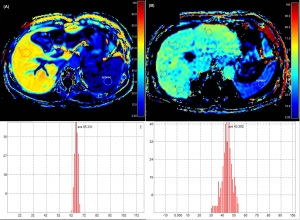
Quantitative MR Provides Liver Function Information in Chronic Liver Disease: Hepatocyce fraction derived from gadoxetic acid enhanced MRI correlates to a commonly used marker for chronic liver disease. #910, Room 311, 16:15
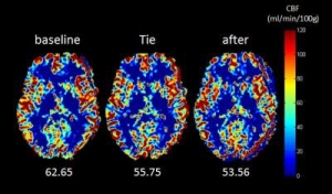
Relax the Dress-Code! #2579 demonstrates that wearing a necktie tightly can affect cerebral blood flow as measured with pCASL MRI

Scott’s Corner
Fantastic meeting so far. Come see the Special Session – Scientific Highlights of the 25th Annual Meeting during the Plenary session.
Scott Reeder, 2017 Program Chair
Karla’s Corner
Today’s ISMRM is all about computation! A day full of Machine learning – morning scientific session, Junior Fellows Symposium at 13:45, followed by a discussion in The Resonarium at 5pm. Also in The Resonarium: Secret Sessions on algorithm development and the BART environment.
Karla Miller, 2017 Education Chair

