By Blake Dewey

Bernhard Strasser
This week we gathered across multiple continents (as I feel we always do!) to discuss the ins and outs of spectroscopic imaging with Bernhard Strasser and Wolfgang Bogner, two authors of “(2 + 1)D-CAIPIRINHA accelerated MR spectroscopic imaging of the brain at 7T”, one of the MRM Editor’s Picks for August 2017. In this paper, Bernhard and his colleagues propose a new method of acceleration for magnetic resonance spectroscopic imaging (MRSI) that combines 2D CAIPIRINHA with simultaneous multi-slice (SMS) to accelerate imaging in all three spatial dimensions.
Blake: Let’s begin with a bit of background. How did you become interested in the field of MRI/MRSI?
Bernhard: I just started about 6 or 7 years ago. I began being interested in fMRI and how the brain works and what happens when we are afraid or we see something pleasant. It turned out that I found MRI spectroscopy to be even more interesting.
Wolfgang: My background is in physics and after my Master’s thesis I became bored of radiation prediction, but wanted to stay in the life sciences. I found MR interesting when I came across it in my studies and started my PhD in the field. I have pretty much worked with brain spectroscopy from the beginning.
Blake: Now moving on to the paper, can you give a brief summary of this work?
Bernhard: What we did in this paper was to create a new method using parallel imaging to accelerate the acquisition of MRSI in all three of the spatial dimensions. We do 2D CAIPIRINHA in-plane and then because we have multiple slices, we do simultaneous multi-slice (SMS) in the slice direction. We then compare this to more common parallel imaging methods, 2D GRAPPA and 2D CAIPIRINHA.
Blake: Does this require more intricate measurement of sensitivities or can we use a standard method for calculating the required weights?
Bernhard: We can use a standard GRAPPA technique. The only difference is we collect an imaging sequence, a gradient echo sequence, to gather the calibration data. We don’t do this with the spectroscopy data, because it would take too much time.
Wolfgang: Our pre-scan also does not include water suppression which gives us many times more signal to use for calculation of GRAPPA or CAIPIRINHA weights. They are pretty much noiseless. These can be then directly applied to the spectroscopic acquisitions.
Blake: What would you say is the biggest strength of this method?
Bernhard: I would say that the ability to accelerate in all 3 dimensions. That way we are exploiting the sensitivities in each direction and that is very beneficial. Also, the way we acquire the calibration data, but this is just one puzzle piece of our whole methodology. In general, I would say that we can measure, within a reasonable measurement time, very high resolution MRSI data with very high SNR and I think there is a lot of information that can be extracted from this data. Unfortunately MRSI is not often used clinically, but I think this would be a big benefit of our method, as we were even able to use it in a clinical study.

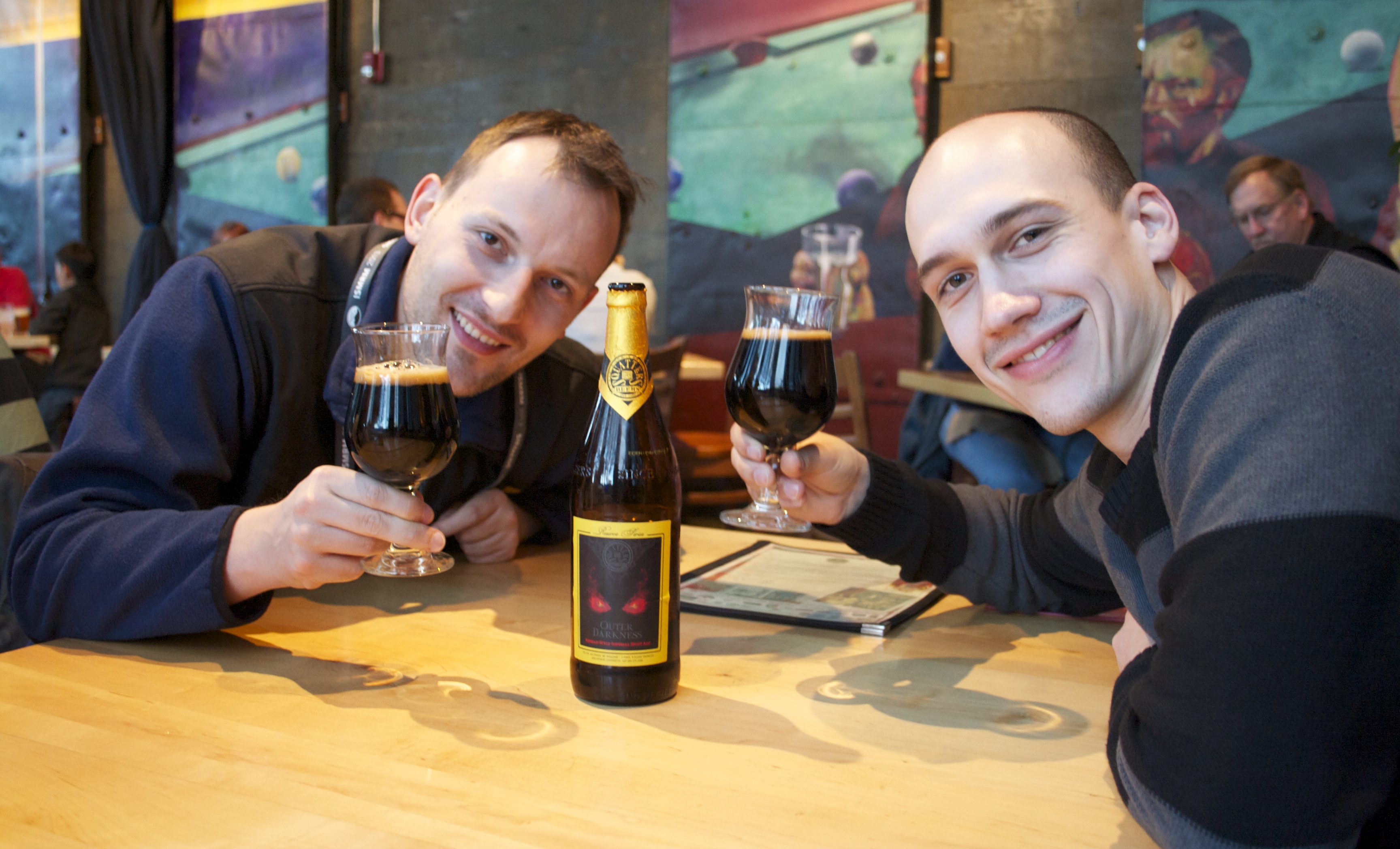
Bernhard Strasser (left) and Wolfgang Bogner (right)
Blake: Tell me more about this clinical study.
Wolfgang: We can show multiple cases of MS patients, where we have 5, 6, 7 different chemical compounds in metabolic maps with a matrix size of 100×100, or slightly above. This has not been possible before. In some cases, where we have very small lesions, on the order of 3mm, we can suddenly see metabolic changes with that resolution. For example, we can observe myo-inositol or NAA in lesions that are only about 2-3 mm large.
Blake: What would you say the biggest weakness of this method is?
Bernhard: The most difficult part is determining the best patterns, the 2D CAIPIRINHA patterns plus the FOV shift from the SMS. Initially, I thought it is very straightforward, you just test all of the possible patterns, but the problem is there are so many possible patterns, that takes quite some effort. Also, whenever you change, for example, the scanner or the coil, you would need to find the best patterns again.
Blake: Do you optimize the patterns on each subject or do you do it on each coil and attempt to position the patient similarly?
Bernhard: The latter. It shouldn’t change too much from patient to patient as long as the brains or the heads are not so different. What is important is how the sensitivity map looks, this influences the patterns, because of the aliasing that we achieve with the (2+1)D CAIPIRINHA.
Blake: What is the next step for this research?
Bernhard: I am working on accelerating MRSI acquisition even more, using spectral-spatial encoding, where spatial and spectral information is encoded at the same time using spirals or EPSI. This will help us get bigger coverage in the z direction to cover even more of the brain.
Wolfgang: Although Bernhard has recently moved from Vienna to Boston, we are still working together, with some of the same common goals. We basically want to have 3D whole brain coverage for spectroscopic imaging and have it in clinical use. MS is where our clinical collaborations have worked very well and we already have 70 scans performed. These clinical scans are only done with the methods that are described in this paper because we know that these sequences are robust. In parallel we are developing spectral-spatial encoding techniques and only when this runs with 3D and is really robust with scanner reconstruction, will we be able to use this in clinical studies.
Blake: Thank you so much for speaking with us today. Is there anything else you would like to add?
Wolfgang: We are always looking for collaborators that are willing to try out our methods. We also have a reconstruction pipeline that we are eager to share, if anyone is interested. Thanks so much.
