 The first Editor’s Pick of 2016 is from Hye-Young Heo and Jinyuan Zhou, researchers at John Hopkins University. Their paper presents a new method, Extrapolated semi-solid Magnetization transfer Reference (EMR), which quantitatively measures two subsets of Chemical Exchange Saturation Transfer (CEST): Amide Proton Transfer (APT) and Nuclear Overhauser Enhancement (NOE). They tested the EMR method in an animal glioma tumor model, and were able to distinguish between active tumor regions, necrotic regions, and healthy tissue. They also pointed out that their quantitative measure of APT was not confounded by NOE effects. We recently spoke with Hye-Young and Jinyuan about their project, what lead them to APT imaging, and what advice they have for newcomers.
MRMH: Please tell us about yourselves and your background.
Hye-Young: My background is in biomedical engineering, and I did proton exchange-based MRI, T1 rho imaging, for my PhD at the University of Iowa from 2008 to 2013. I joined Dr. Zhou’s group in fall 2013, where I have worked on another proton exchange-based MRI method, CEST imaging.
Jinyuan: I’m an associate professor of Radiology at John Hopkins University, and my background is in MR physics. I got my PhD degree from the Wuhan Institute of Physics at the Chinese Academy of Science in 1996, where I did solid-state NMR. We started doing CEST MRI here around the year 2000.
MRMH: When did you move to the US?

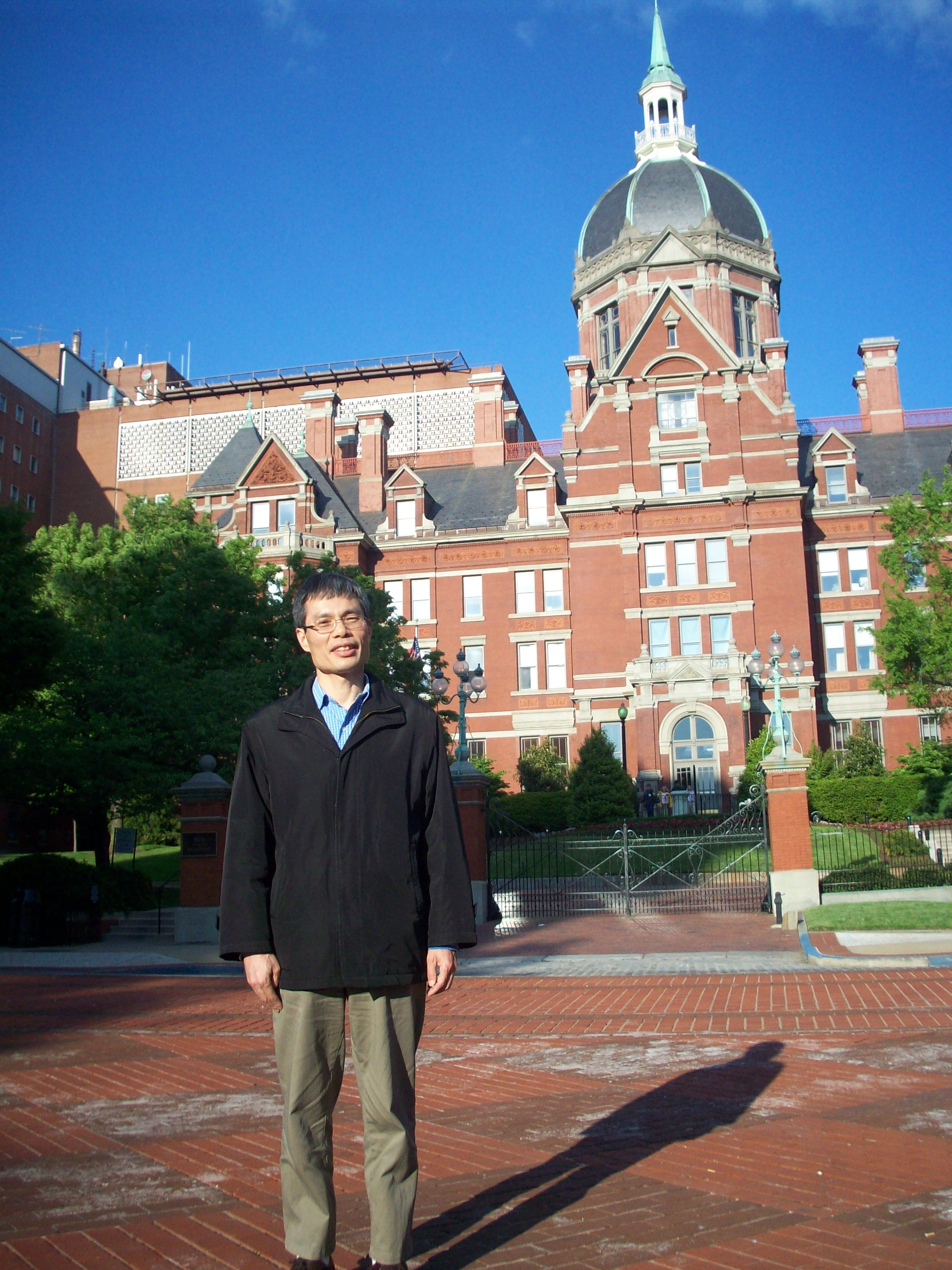
Jinyuan on the beautiful Hopkins campus
Jinyuan: 1997… it’s been almost 18 years [chuckles].
MRMH: What sparked your interest in chemical exchange and magnetization transfer imaging?
Hye-Young: APT imaging, a subset of CEST, is a very interesting topic. It’s an important molecular MRI technique which can detect very low concentrations of amide protons. Clinical applications include tumor detection, treatment assessment, and stroke imaging.
Jinyuan: When I first arrived here, I was a post-doc under the supervision of Dr. Peter van Zijl. Peter has many new ideas and always pushed us to try new things. We started with CEST, which was new at the time, and then that led me to APT. Most MRI techniques are water-based, while APT is protein-based. We don’t have to inject any contrast agents; we just use the endogenous proteins in our body.
MRMH: Could you please give us a quick overview of your paper?
Hye-Young: In general, CEST is confounded by the water direct saturation effect and other magnetization transfer effects. So, in MTR asymmetry quantifications, the CEST signal is confounded by the Nuclear Overhauser Effect (upfield from the water). In this study (together with another paper, DOI: 10.1002/mrm.25795), we introduced a new method called Extrapolated Semi-Solid Magnetization Transfer Reference (EMR), to quantify the pure APT signal by isolating it from confounding factors.
Jinyuan: Usually, using MTR asymmetry, the obtained APT-weighted intensity values range between 2% and 3%. One thing we found with EMR is that the pure APT effect is very large. It can be more than 10% in tumor. It’s very big! Another discovery was that the dominant APT-weighted contrast between tumors and normal tissue is APT, not NOE.
MRMH: For those of us unfamiliar with cancer biology, could you please explain why you observed different exchange phenomena between healthy tissue and tumor (center vs. rim)?
Hye-Young: We scanned the animal tumor model, human glioblastoma-bearing rats, 45 days post-implantation. At this time, the tumor center had begun necrosis, so there were less mobile APT-detectable proteins there. However, the tumor rim is always very active; there are a lot of mobile proteins compared to normal tissue and the tumor center.
MRMH: What advice can you offer to a graduate student who has read your paper and wants to implement EMR in her/his project?
Hye-Young: The idea behind the EMR method is very simple, but there is a lot of complex mathematics in the paper. I would recommend to do Bloch equation-based simulations first, to examine how the CEST signal changes with RF power, T1 and T2 relaxation times, and other experimental settings.
Jinyuan: I think it’s very important that you spend time to optimize your sequence. In my experience, egg white makes a very good/cheap phantom for APT, because it has many natural proteins. Water is not a good phantom for APT. And you should first make sure that your z-spectrum is very smooth, not noisy.
MRMH: Do you lose the APT effect if you cook the egg?
Jinyuan: [laughs] Yes, if the egg is cooked, you can see the APT effect reduces almost to 0.
MRMH: What other topics currently excite you?
Hye-Young: Recently, I’m very interested in fast CEST imaging, using parallel MRI, k-t acceleration, and compressed sensing techniques because the CEST imaging has a relatively long acquisition time due to acquiring multiple RF saturation frequencies. I think fast CEST imaging is great for the evaluation of acute stroke patients and pediatric patients.
Jinyuan: I’m currently particularly interested in radiogenomics. People are doing radiogenomics to find the correlation between MR features and genes (e.g. gadolinium enhancement, FLAIR hyperintensity). I think that APT-weighted MRI features might be more associated with the genome, because APT is protein-based.
MRMH: Thank you and good luck with your future work!
*Interview conducted by Mathieu Boudreau and Nikola Stikov.
